
3D digital holographic microscopic water quality detection system
Abstract:
Water is essential for life, but accessing clean water is a challenge in many places due to pollution and how water is naturally distributed across the Earth. This study evaluates the importance of checking water quality, especially with more factories and cities growing. In the study, we focus on plankton, tiny water organisms that can reveal a lot about water's health. When water gets polluted, the type and amount of plankton change, which is a warning sign of poor water quality. Traditional ways of studying plankton involve collecting water samples and analyzing them in labs, which takes a lot of time and can delay getting important information. To make this process faster and more accurate, we use new technologies like digital holographic microscopy (DHM) and lensless digital holographic microscopy (LDHM). These methods allow us to see plankton in great detail and quickly understand water quality.We explain how we create and analyze images of plankton without using traditional lenses, using light and computers to capture and study these images in detail. This process includes using different light wavelengths and advanced computing methods like the Gerchberg-Saxton (G-S) iteration for better image analysis, and techniques like level-set-based cell image segmentation for counting and studying plankton cells accurately. Our experiments with these technologies allowed us to see tiny details in plankton, as small as 2.46 micrometers, which approaches the upper limits of what our equipment can do. This proves that our approach is effective for quickly checking water quality by looking at plankton.
By introducing these new technologies, we provide a faster and cheaper way to monitor water quality, which could help in better understanding and protecting aquatic environments. This research offers a new solution for quickly assessing water quality through a detailed study of plankton, showing a way forward for environmental monitoring.
Bibliography/Citations:
1. Li, J., Zhou, N., Sun, J. et al. Transport of intensity diffraction tomography with non-interferometric synthetic aperture for three-dimensional label-free microscopy. Light Sci Appl 11, 154 (2022). https://doi.org/10.1038/s41377-022-00815-7
2. M.K. Kim, Digital Holographic Microscopy (Springer, 2011).
3.U. Schnars and W. Jueptner, Digital Holography (Springer, 2005).
4. B. Kemper and G. von Bally, “Digital holographic microscopy for live cell applications and technical inspection,” Appl. Opt. 47(4), A52–A61 (2008).
5. M. Takeda, H. Ina, and S. Kobayashi, “Fourier-transform method of fringe-pattern analysis for computer-based topography and interferometry,” J. Opt. Soc. Am. 72(1), 156–160 (1982).
6. M. Pirga and M. Kujawinska, “Two directional spatial-carrier phase-shifting method for analysis of crossed and closed fringe patterns,” Opt. Eng. 34(8), 2459 (1995).
7. T. Ikeda, G. Popescu, R. Dasari, and M. Feld, “Hilbert phase microscopy for investigating fast dynamics in transparent systems,” Opt. Lett. 30(10), 1165–1167 (2005).
8. Y. Park, C. Depeursinge, and G. Popescu, “Quantitative phase imaging in biomedicine,” Nat. Photonics 12(10), 578–589 (2018).
9. T. Kozacki and K. Falaggis, “Angular spectrum-based wave-propagation method with compact space bandwidth for large propagation distances,” Opt. Lett. 40(14), 3420–3423 (2015).
10. Y. Zhang, H. Wang, Y. Wu, M. Tamamitsu, and A. Ozcan, “Edge sparsity criterion for robust holographic autofocusing,” Opt. Lett. 42(19), 3824–3827 (2017).
11. Y. Wu, Y. Rivenson, Y. Zhang, Z. Wei, H. Günaydin, X. Lin, and A. Ozcan, “Extended depth-of-field in holographic imaging using deep-learning-based autofocusing and phase recovery,” Optica 5(6), 704–710 (2018).
12. A. Ozcan and E. McLeod, “Lensless Imaging and Sensing,” Annu. Rev. Biomed. Eng. 18(1), 77–102 (2016).
13. E. McLeod and A. Ozcan, “Unconventional methods of imaging: computational microscopy and compact implementations,” Rep. Prog. Phys. 79(7), 076001 (2016).
14. D. Gabor, “A New Microscopic Principle,” Nature 161(4098), 777–778 (1948).
15. T. Agbana, H. Gong, A. Amoah, V. Bezzubik, M. Verhaegen, and G. Vdovin, “Aliasing, coherence, and resolution in a lensless holographic microscope,” Opt. Lett. 42(12), 2271–2274 (2017).
16. Vashist SK, Mudanyali O, Schneider EM, Zengerle R, Ozcan A. 2013. Cellphone-based devices for bioanalytical sciences. Anal. Bioanal. Chem. 406:3263–77
17. Ozcan A. 2014. Mobile phones democratize and cultivate next-generation imaging, diagnostics and measurement tools. Lab Chip 14:3187–94
18. Greenbaum A, Ozcan A. 2012. Maskless imaging of dense samples using pixel super-resolution based multi-height lensfree on-chip microscopy. Opt. Expr. 20:3129–43
19. Bishara W, Sikora U, Mudanyali O, Su T-W, Yaglidere O, et al. 2011. Handheld, lensless microscope identifies malaria parasites. SPIE Newsroom, Aug. 5. http://spie.org/x51571.xml
20. Su T-W, Choi I, Feng J, Huang K, McLeod E, Ozcan A. 2013. Sperm trajectories form chiral ribbons. Sci. Rep. 3:1664
21. Seo S, Isikman SO, Sencan I, Mudanyali O, Su T-W, et al. 2010. High-throughput lens-free blood analysis on a chip. Anal. Chem. 82:4621–27
Additional Project Information
Research Plan:
Question:
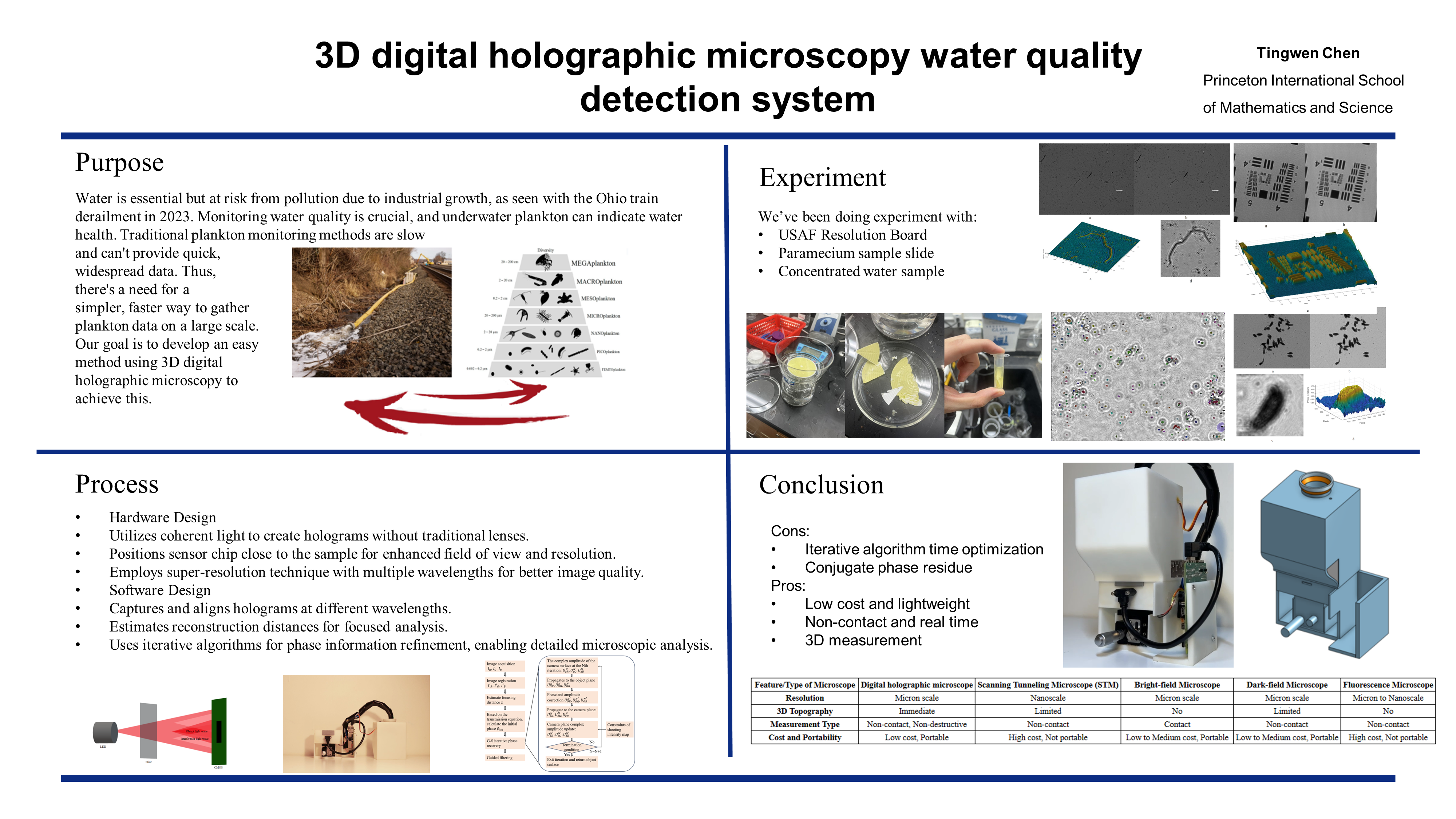
Water is a non-renewable resource that human society depends on for survival. However, with the continuous advancement of industrialization and urbanization, the pollution of limited water resources is also facing an increasingly serious risk. For example, on February 3, 2023, a train derailment in Ohio released toxic chemicals into the environment, causing significant water contamination for local residents. Therefore, the detection of water quality has become particularly important. Underwater plankton is closely related to the quality of the corresponding living water environment, through the detection of underwater plankton species, physiological composition changes, life activity patterns and quantity, and other characteristics, can indirectly or directly obtain information on the quality of the water body and the trend of changes in the quality of the water body. Most of the current detection methods for underwater plankton are based on observations. However, when the sampling is completed and then put into the laboratory for research, the experimental results obtained have a great delay, and it is difficult to obtain large-scale experimental data in time, so it is not possible to carry out timely and effective monitoring and evaluation of the water quality pollution situation. How to use relatively simple operations can be obtained in time and space on a large scale effective underwater plankton data is an important research direction. So our goal here is to develop a simple structure that could easily obtain the underwater plankton data using 3D digital holographic microscopic technology.
Goal:
1. How to detect plankton based on digital holographic microscopy and determine water quality based on this.
2. To explore the imaging mechanism of digital holographic microscopy.
3. How to design a low-cost and portable digital holographic microscopic prototype to achieve rapid 3d imaging of underwater plankton, and consider how to model the instrument by 3D printing.
4. How to develop an algorithm based on multi-wavelength digital holographic microscopy to realize the phase imaging of plankton
5. Conduct experiment to test the performance of the system under the requirement of water quality detection.
Expected outcome:
By using our structure and algorithm, with inexpensive hardware, the resolution of the 3D microscope could be greatly improved. After conducting experiment based on water quality detection, a machine learning algorithm could be developed so that the water quality detection could be easily done by analysing the data of algae captured by the device.
Hypotheses:
Using different optic filter on the white led, it’s possible to implement an phase recovery algorithm based on multi-wavelength to the 3D digital holograph microscope. Based on processed picture, the algae data of the sample water could be collected and analysed thus the water quality could be evaluated.
Procedures:
- Research on the background of detecting plankton based on digital holographic microscopy technology to detect water quality.
- Investigation of the relationship between water quality and plankton.
- A survey of current plankton detection techniques.
- Research on plankton detection based on the principle of digital holography.
- The basic principle of digital holographic microscopy, algorithm simulation, and implementation.
- Algorithm simulation of digital holographic interference recording.
- Algorithm simulation for diffraction reproduction of digital holography.
- Digital Holographic 3D Imaging and Morphological Analysis Based on Machine Learning.
- Hardware design and construction based on holographic structure.
- 3d modeling design of digital holographic microscopic prototype based on onshape platform.
- Purchase the necessary equipment, including white LED, Basler dart daA2500, SG90 micro servo, 650nm,532nm,450nm narrow band filters, micro vertical displacement stage, and Raspberry Pi 4 Model B, and complete the relevant instrument test.
- 3d printing of shell support parts based on structure design.
- Equipment assembly and system debugging based on structure design.
- Based on the test feedback, replace the parts and adjust the 3d model, and finally complete the hardware design of the digital holographic microscopy system.
- Develop and test of the algorithm
- Phase retrieval algorithm for in-line digital holography based on GS iteration at single wavelength
- Phase retrieval algorithm for in-line digital holography based on multi-height GS iteration
- Based on Raspberry PI microcontroller and related hardware, develop the wireless control program of digital holographic microscopy hardware system.
- Phase retrieval algorithm for in-line digital holography based on multi-wavelength GS iteration
- The combination of software and hardware is carried out through preliminary experiments.
- Place the sample on the holder in front of CMOS, keeping the distance as close as possible.
- Wirelessly connect to the Raspberry PI and send analog signals to control the servo turns specific angle, carrying the plate turns to match specific optic filter.
- Recording the information from the CMOS by connect CMOS to Raspberry PI and using Pylon Viewer to record the image, then send the set of image back to the contral computer to analysis.
- Redo the experiment 2 times using different optic filter.
- Conduct experiments and data analysis.
- Purchase plankton samples for performance testing of the equipment
- Collect water samples near the experimental site and analyze plankton samples in the field
- Obtain the phase information of the sample by image processing based on the collected plankton information of the sample
- Machine learning-based morphological analysis based on phase information of samples
Questions and Answers
1. What was the major objective of your project and what was your plan to achieve it?
Water is a non-renewable resource that human society depends on for survival. However, with the continuous advancement of industrialization and urbanization, the pollution of limited water resources is also facing an increasingly serious risk. For example, on February 3, 2023, a train derailment in Ohio released toxic chemicals into the environment, causing significant water contamination for local residents. Therefore, the detection of water quality has become particularly important. Underwater plankton is closely related to the quality of the corresponding living water environment, through the detection of underwater plankton species, physiological composition changes, life activity patterns and quantity, and other characteristics, can indirectly or directly obtain information on the quality of the water body and the trend of changes in the quality of the water body. Most of the current detection methods for underwater plankton are based on observations. However, when the sampling is completed and then put into the laboratory for research, the experimental results obtained have a great delay, and it is difficult to obtain large-scale experimental data in time, so it is not possible to carry out timely and effective monitoring and evaluation of the water quality pollution situation. How to use relatively simple operations can be obtained in time and space on a large scale effective underwater plankton data is an important research direction. So our goal here is to develop a simple structure that could easily obtain the underwater plankton data using 3D digital holographic technology.
To achieve this goal, the whole process is broken into six steps
1)Research about the relationship between algae and water quality.
2)Research about the requirements of the device such as resolution or function.
3)Hardware design and construction based on holographic structure.
4)Software debugging and algorithm development.
5)The combination of software and hardware is carried out through preliminary experiments.
6)Conduct experiments and data analysis.
2.What were the major tasks you had to perform in order to complete your project?
It is very difficult to build a lightweight and inexpensive holographic microscopic system. During my research, I had to design the overall 3D holographic microstructure and complete the assembly of the prototype. The code for the entire system also needed to be debugged and experimented with to ensure the accuracy of the results captured by the system
3.What is new or novel about your project?
1)In this research, we are trying to use algae as an indicator of water quality
2)During the designing of the structure, we are trying to build a lightweight and inexpensive microscopic system, where most of the microscopic systems have become more and more cumbersome, complex, challenging to maintain, and increasingly expensive.
3)During the designing of the structure, we developed a system using a light filter and servo to generate lights with different wavelengths, to increase the accuracy of the result captured by the system. By reading relevant references and browsing mainstream production companies, we didn't find a similar structure.
4. What was the most challenging part of completing your project?
When building up the structure of the device, since our goal is to make the device lightweight and inexpensive, the space is very limited, making the maintenance and modification of the device very difficult. Apart from the design of the structure, all the transparent surfaces in the lightpath need to be clean to ensure the quality of the image captured. Because of the closed system, it’s difficult to clean the surface after assembling the system. Therefore, after the first prototype had been built, I changed the structure so that the whole system could be easily assembled and dismantled.
During this experience, I learned that when doing a precise experiment or building up a precise device, all the possible problem and their solution should be carefully considered. Every part of the design is important to the success of the experiment.
5. If you were going to do this project again, are there any things you would you do differently the next time?
I’ll try to build up a system that is waterproof so that the whole system can be implemented underwater so that the quality of the water can be monitored in real-time.
6. Did working on this project give you any ideas for other projects?
1)It’s possible to implement a similar structure to the air quality monitor system since the pollution particles in the air are about the size of a pixel, which means that it’s possible to detect them using a similar structure.
2)It’s possible to create a 3D structure for the cell observed using a similar system.
7. How did COVID-19 affect the completion of your project?
No